
Tomosynthesis, often called 3D mammography, usually gives doctors a clearer look at breast tissue than traditional 2D mammography. It is not a different goal; it is a different way of seeing. For many people, especially those with dense breasts, that extra detail can mean fewer repeat appointments and a better chance of spotting small cancers early.
TLDR: 3D mammography takes multiple low-dose X-ray images and builds thin “slices” of the breast, while traditional mammography creates flat 2D views. In studies, tomosynthesis has been linked with about 15% to 30% fewer callbacks and can find roughly 1 to 2 more cancers per 1,000 screenings compared with 2D alone. For example, if 1,000 women have screening mammograms and 100 would have been called back after 2D imaging, 3D imaging may reduce that number by 15 to 30 people. It still uses compression, and it still is not perfect, but it can make breast imaging less stressful and more precise.
What Traditional Mammography Shows
Traditional mammography is also called 2D mammography. It takes X-ray images of the breast from two main angles. These are usually a top-to-bottom view and a side-angle view.
The breast is compressed between two plates. Yes, that part is uncomfortable. It can feel awkward and irritating, even when the technologist is skilled and kind. The compression spreads out the tissue, reduces blur, and lowers the radiation dose needed for a sharp image.
In a 2D mammogram, all the tissue is shown in a flat image. This is useful, but it has a known problem. Breast tissue can overlap. A normal area may look suspicious because tissue stacks on top of itself. A small cancer may also hide behind dense tissue.
That overlap is one reason some people are asked to return for more images. The callback does not always mean cancer. Often, it means the radiologist needs a cleaner view. Still, waiting for that answer can be stressful.
What Tomosynthesis Adds
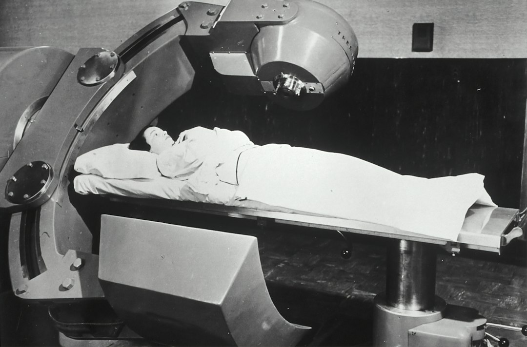
Tomosynthesis is commonly called 3D mammography, though it is not a hologram or a full 3D model like a CT scan. During the exam, the X-ray tube moves in an arc over the breast. It captures many low-dose images from different angles.
A computer then rebuilds those images into thin layers. A radiologist can scroll through them, almost like flipping through the pages of a book. Instead of seeing all tissue piled into one flat picture, the doctor can inspect one layer at a time.
This can help with two big issues:
- Finding small cancers that may be hidden in overlapping tissue.
- Reducing false alarms caused by normal tissue that only looks suspicious on a flat image.
Honestly, it feels like breast imaging should have worked this way sooner. A flat image of a rounded, layered body part can only tell part of the story. Tomosynthesis gives the radiologist more context.
3D Mammography vs 2D Mammography
The main difference is depth. Traditional mammography gives a flat view. Tomosynthesis gives a layered view.
| Feature | Traditional 2D Mammography | 3D Mammography Tomosynthesis |
|---|---|---|
| Image type | Flat images | Thin image slices |
| Tissue overlap | More overlap | Less overlap |
| Callback rate | Often higher | Often lower |
| Use with dense breasts | Helpful, but limited | Often more helpful |
| Exam feel | Compression required | Compression required |
The experience for the patient is very similar. You stand at the machine. The breast is compressed. Images are taken. The whole exam often takes about the same time, though 3D image capture may add a few seconds per view.
The reading process is different. Radiologists review more images with tomosynthesis. That can take longer on their end. The catch is that more data can also mean more confidence, which may save patients from extra imaging later.
Why Dense Breast Tissue Matters
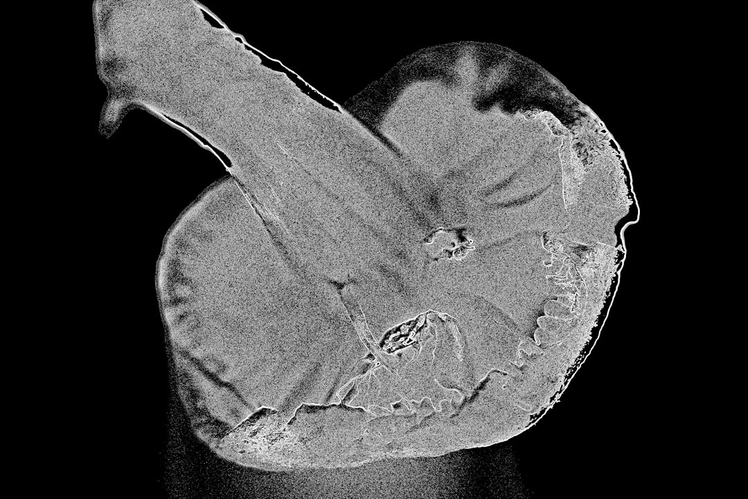
Dense breast tissue is common. It means the breast has more fibrous and glandular tissue and less fatty tissue. On a mammogram, dense tissue appears white. Many cancers also appear white. That makes finding cancer harder.
Think of it like looking for a snowball in a snowstorm. The object is there, but the background makes it tough to see.
Tomosynthesis can help because it separates tissue into slices. It does not make dense breasts transparent. It does not replace every other test. But it may improve visibility enough to change what the radiologist can detect.
Some people with dense breasts may also need ultrasound or breast MRI, depending on personal risk. That decision depends on factors such as age, family history, genetic risk, prior biopsies, and past cancer history.
Does 3D Mammography Find More Cancer?
In many screening programs, tomosynthesis finds slightly more breast cancers than 2D mammography alone. The increase is not huge, but it matters. Finding even one extra early cancer per 1,000 screenings can affect real lives.
Tomosynthesis is especially good at finding invasive cancers, which are cancers that have moved beyond the milk ducts or lobules into nearby breast tissue. These are often the cancers doctors most want to catch early.
Traditional mammography is still strong at detecting calcifications. These are tiny calcium deposits that may signal early breast changes, including ductal carcinoma in situ, also called DCIS. For this reason, many centers use 3D imaging along with a 2D image or a computer-generated synthetic 2D image.
What About Radiation?
Both 2D and 3D mammography use low-dose X-rays. Radiation exposure from mammography is small and regulated. For most people, the benefit of finding breast cancer early outweighs the radiation risk.
When 3D mammography first became common, some exams used both 2D and 3D images, which raised the dose slightly. Many modern systems now create a synthetic 2D image from the 3D data. This can keep the dose closer to standard mammography levels.
If you are concerned, ask the imaging center what type of system they use. A simple question works: “Will my exam include synthetic 2D images, standard 2D images, or both?”
Who May Benefit Most from Tomosynthesis?
3D mammography can be useful for many screening patients, but some groups may benefit more than others.
- People with dense breasts, because overlapping tissue can hide findings.
- People who have had callbacks before, especially for tissue overlap.
- Patients having a diagnostic mammogram for a lump, pain, nipple changes, or prior abnormal imaging.
- People at average risk who want the most detailed standard screening option available.
Here is a simple user case. Maria, 47, has heterogeneously dense breasts. Her last 2D mammogram led to a callback, extra images, and a week of worry. The finding was normal tissue overlap. The next year, she has 3D mammography. The radiologist can scroll through the layered images and sees the area clearly. No callback is needed.
Image not found in postmetaLimitations and Costs
Tomosynthesis is helpful, but it is not magic. It can still miss cancers. It can still find spots that need extra imaging. It may cost more, depending on the facility and insurance plan.
Coverage has improved in many regions, but billing can still be annoying. Expect to waste time on a phone call if your plan is vague about “screening mammography” versus “3D mammography.” Before the appointment, ask the imaging center for the exact billing code and check with your insurer.
Availability also varies. Large hospitals and breast centers often have 3D systems. Smaller centers may not. That does not mean 2D mammography is poor care. A high-quality 2D mammogram read by an experienced breast radiologist remains a valuable screening tool.
Which Should You Choose?
If 3D mammography is available and covered, many patients choose it because it can reduce callbacks and improve detection. This is especially reasonable if you have dense breasts or a history of unclear mammograms.
If only 2D mammography is available, do not skip screening. Regular mammography is still one of the best tools for finding breast cancer early. The best test is the one you can access, afford, and repeat on schedule.
Ask three questions before your next breast imaging appointment:
- Do I have dense breast tissue?
- Is 3D mammography available for my screening?
- Will my insurance cover it as preventive care?
Tomosynthesis gives radiologists a sharper way to examine layered breast tissue. Traditional mammography still matters, but 3D imaging often offers a clearer view with fewer gray areas. For many patients, that means less waiting, fewer repeat images, and a better shot at early answers.